Staging reference

Prostate Cancer T-Staging

What is palpable, what MRI shows, and why local extent matters

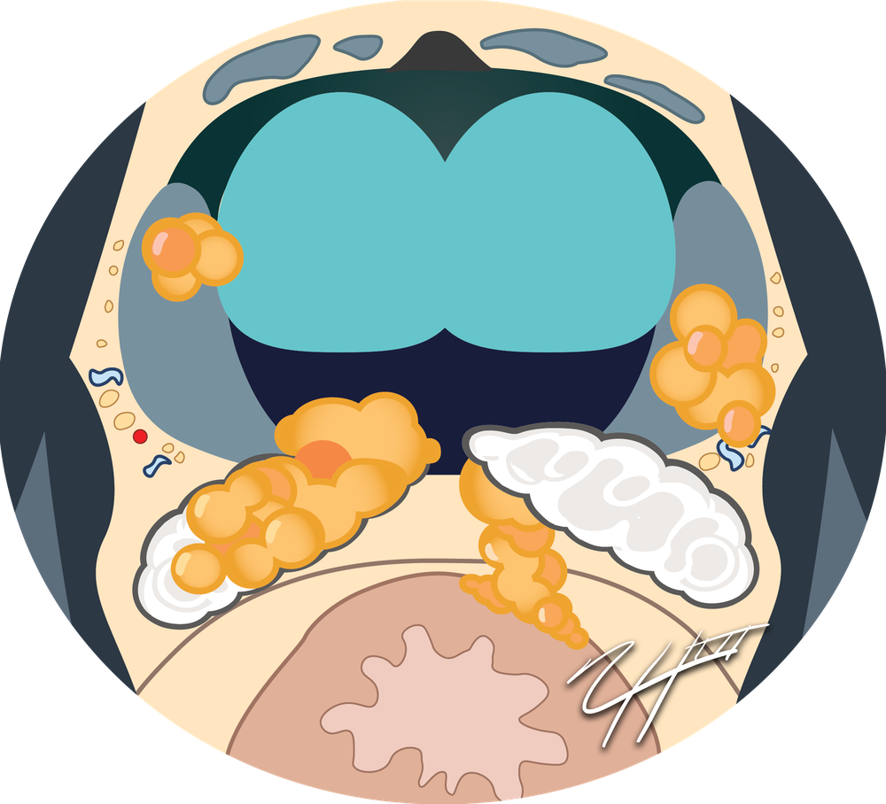
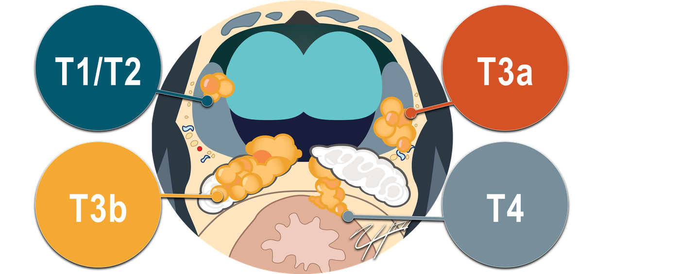
Axial illustration of the prostate with tumor foci confined to the gland, extending beyond the capsule, into the seminal vesicle, and toward the rectum

T Stage at a Glance

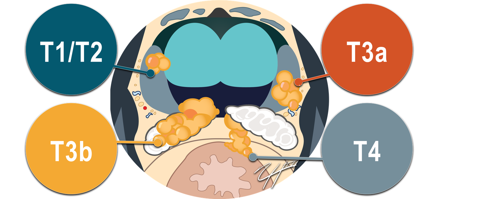
Axial prostate illustration labeled T1/T2 for tumor confined to the gland, T3a for extraprostatic extension, T3b for seminal vesicle invasion, and T4 for invasion of adjacent structures
Hover or select a stage · Tap image to enlarge
Key teaching point

Clinical T Stage ≠ MRI Local Stage

c = clinicalp = pathologic
cT1c = clinical T stage 1c
First c · clinical prefixFinal c · T1 subcategory

Clinical T stage is based on DRE. MRI local extent is reported separately.

Example
DRENo palpable tumor
BiopsyProstate cancer
MRIDefinite extraprostatic extension
Result Clinical stage: cT1c MRI: Findings of extraprostatic extension
Advertisement
T1

Not Palpable

T1aIncidental tumor in ≤5% of resected tissueExample: Small focus of cancer discovered after TURP
T1bIncidental tumor in >5% of resected tissueExample: Cancer involving 10% of TURP tissue
T1cTumor identified by needle biopsy but not palpableExample: Elevated PSA → positive biopsy → normal DRE

MRI size or bilaterality does not automatically make a nonpalpable tumor cT2.

T2

Palpable but Confined

T2a≤ half of one lobe
T2b> half of one lobe, but not both lobes
T2cBoth lobes

Clinical T2 subdivision is based on examination, not MRI tumor size or bilaterality.

T3a

Extraprostatic Extension

Clinical definition (cT3a)Palpable extension through the prostate capsule
MRI local extentAssess and report extraprostatic extension separately
MRI clues
  • Capsular irregularity or disruption
  • Tumor extending into periprostatic fat
  • Measurable extraprostatic tumor
  • Neurovascular bundle involvement
  • Obliteration of the rectoprostatic angle
Example

Peripheral zone tumor with definite extension beyond the prostatic margin

Why it matters

May affect recurrence risk, surgical margins, nerve-sparing decisions, and radiation planning.

T3b

Seminal Vesicle Invasion

Clinical definition (cT3b)Seminal vesicle invasion in the DRE-based category
MRI local extentAssess and report seminal vesicle invasion separately
MRI clues
  • Direct tumor extension from the prostate
  • Low T2 signal replacing normal seminal vesicle architecture
  • Restricted diffusion within the involved seminal vesicle
  • Loss of the normal prostate-seminal vesicle angle
Example

Tumor at the prostate base directly extends into the ipsilateral seminal vesicle

Why it matters

Represents more advanced local disease and affects prognosis and treatment planning.

T4

Adjacent Structure Invasion

Clinical definition (cT4)Fixed tumor or invasion of adjacent structures other than seminal vesicles
MRI local extentAssess and report adjacent-structure invasion separately
External sphincter Rectum Bladder Levator muscles Pelvic wall
Example

Tumor extends through the rectoprostatic plane and directly invades the rectal wall

Why it matters

T4 represents locally advanced disease and can substantially alter treatment planning.

Advertisement

What Does MRI Add?

MRI is best at detecting macroscopic local extension.

No visible EPE on MRI does not exclude microscopic EPE.

T Stage Is Only One Part of Risk

PSA
Grade Group
Biopsy findings
Clinical stage
cT1–T2
Clinically localized diseaseRisk can still range from low to high depending on PSA, Grade Group, and biopsy findings.
cT3–T4
Locally advanced diseaseGreater implications for recurrence risk and treatment planning.
Other categories: TX / T0
TXPrimary tumor cannot be assessed
T0No evidence of primary tumor
Quick recall
T1Not palpable
T2Palpable + confined
T3aEPE
T3bSVI
T4Adjacent structure invasion

Test Yourself

01 A patient has prostate cancer diagnosed on biopsy. DRE is normal, but MRI shows definite extraprostatic extension. What is the clinical T stage?

02 On DRE, tumor extends beyond the prostate into the periprostatic tissues without seminal vesicle invasion. What clinical T stage does this correspond to?

03 On DRE, tumor involvement of a seminal vesicle is identified. What clinical T stage does this represent?

04 On clinical examination/DRE, tumor is fixed or there is invasion of an adjacent structure such as the rectal wall. What clinical T stage does this represent?

Related tools and articles

Written by Nicholas H. Shaheen, MD • Last reviewed October 2026

Medical education note: This article is intended for medical education and workflow support only. It should not replace local practice standards, institutional protocols, or formal society guidelines.