Prostate MRI Toolkit 
Interactive prostate MRI workflow support
Practical tools for prostate MRI interpretation.
An educational workspace with calculators, anatomy guides, scoring assistants, and pop quizzes to help make prostate MRI easier to interpret, teach, and learn.
What's New
A radiologist-friendly guide to PI-RADS, PSA density, PRECISE, Prolaris, Decipher, and how prostate cancer risk tools fit together.
Read article → Article DBSI in Prostate MRIA trainee-friendly explanation of diffusion basis spectrum imaging and how it looks beyond the ADC map.
Read article → New PI-QUAL Assistant addedDescriptor-first PI-QUAL v2 output with sequence-quality scoring and limiting-factor documentation.
Open → Updated Anatomy Assistant updated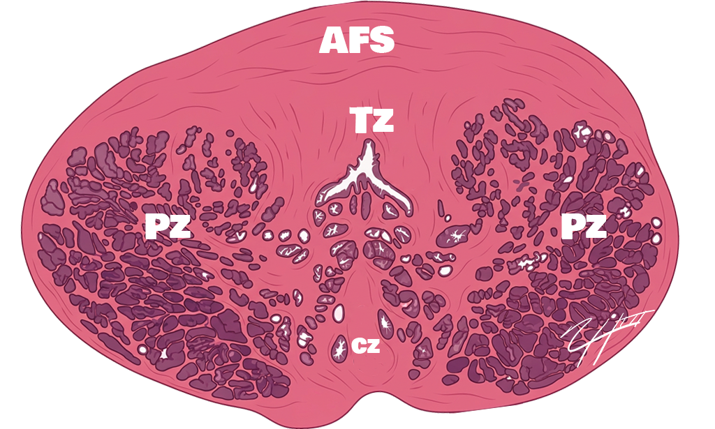
Interactive prostate anatomy guide for zone and localization review.
Explore anatomy → Updated PI-RADS visual decision tree addedA clearer pathway view for peripheral zone and transition zone scoring.
Open decision tree → Updated Monthly Quiz updatedNew case-style prostate MRI questions for trainees and practicing radiologists.
Take the quiz → Updated PRECISE Assistant updatedActive surveillance scoring helper with article link and cleaner result workflow.
Open →PSA Density Pro
Calculate prostate volume and PSA density, then generate a clean report phrase.

PI-RADS Assistant
Descriptor-driven PI-RADS scoring with a concise PI-RADS v2.1 quick guide.
Anatomy Assistant
Review zonal anatomy.

Post-Treatment MRI Assistant
Review post-treatment recurrence assessment patterns for prostate MRI.
PRECISE Assistant
Review follow-up response patterns using PRECISE-style assessment logic.
PI-QUAL Assistant
Assess prostate MRI technical quality using PI-QUAL v2-style logic.
Monthly Quiz
Monthly pop quiz.
References & More
Find core references, source documents, and expanded educational articles.
What is Prostate MRI Toolkit?
Prostate MRI Toolkit is an educational prostate MRI resource created by Nick Shaheen, MD, a board-certified radiologist with fellowship training in abdominal imaging and intervention.
The goal of this website is to provide practical, easy-to-use educational tools for radiologists, trainees, urologists, and healthcare professionals involved in prostate MRI interpretation and prostate cancer evaluation.
The calculators, anatomy modules, and decision-support tools presented throughout this site are based on published prostate MRI literature, PI-RADS guidance, and commonly utilized clinical workflows. References, educational articles, and external resources for the tools included on this website are available on the References & More page.