Prostate Anatomy Overview 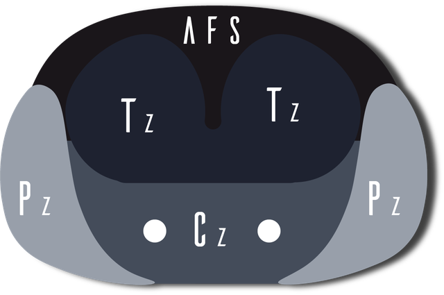
Key anatomic zones and MRI interpretation pearls for prostate MRI.
Peripheral Zone (PZ)
- Largest prostatic zone.
- Approximately 70% glandular tissue.
- Most prostate cancers originate in the peripheral zone, approximately 70%.
- Typically high T2 signal unless replaced by tumor or prostatitis.
Transition Zone (TZ)
- Composed of approximately 60% stromal tissue and 40% glandular tissue.
- Site of benign prostatic hyperplasia.
- Approximately 20% of prostate cancers arise in the transition zone.
- Can appear heterogeneous due to BPH nodules and stromal hyperplasia.
Central Zone (CZ)
- Approximately 25% glandular tissue.
- Only 1–5% of prostate cancers originate in the central zone.
- Typically symmetric and relatively low T2 signal.
- Can occasionally mimic malignancy at the prostate base.
Periurethral Zone & Anterior Fibromuscular Stroma
- Rare site of prostate cancer origin.
- Predominantly fibromuscular tissue with low T2 signal.
- The prostate has no true capsule; the so-called capsule represents a fibromuscular pseudocapsule.
- The pseudocapsule is incomplete anteriorly and at the apex.
Peripheral Zone vs Transition Zone Lesions
Lesion location changes the PI-RADS pathway: peripheral zone lesions are primarily assessed by DWI/ADC, while transition zone and anterior lesions are primarily assessed by T2 morphology. Read the overview of why zone location matters. For a concise scoring refresher, see the PI-RADS v2.1 Quick Guide.
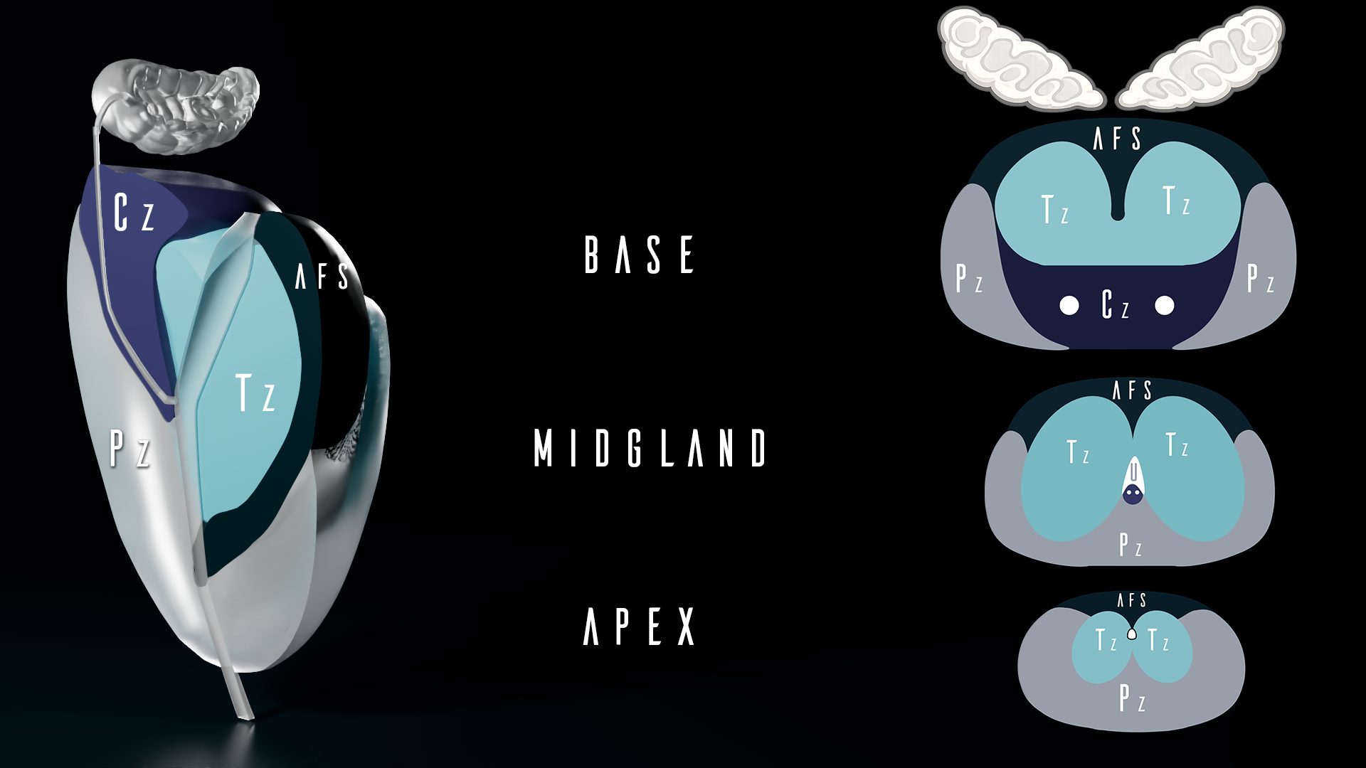
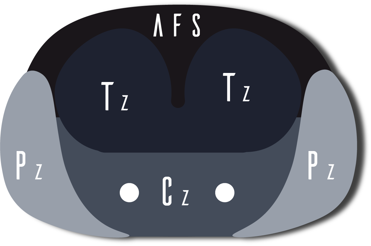
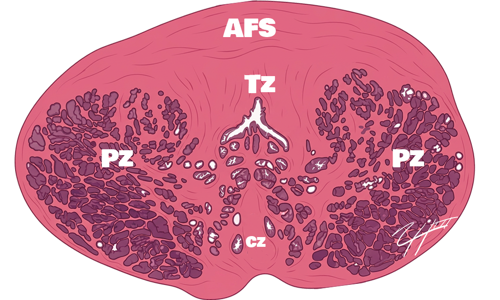
PI-RADS v2.1 Sector Map
PI-RADS v2.1 divides the prostate into the base, midgland, and apex along the craniocaudal axis. Within each level, lesions are localized by side and by anterior-versus-posterior position to create a standardized sector map. The zonal anatomy within those sectors, including the peripheral zone, transition zone, central zone, and anterior fibromuscular stroma, helps further refine lesion location. This framework improves communication, supports targeted biopsy and treatment planning, and makes follow-up comparisons more consistent.