Nodal Staging: Know the Boundary
A visual guide to regional and nonregional lymph nodes in prostate cancer
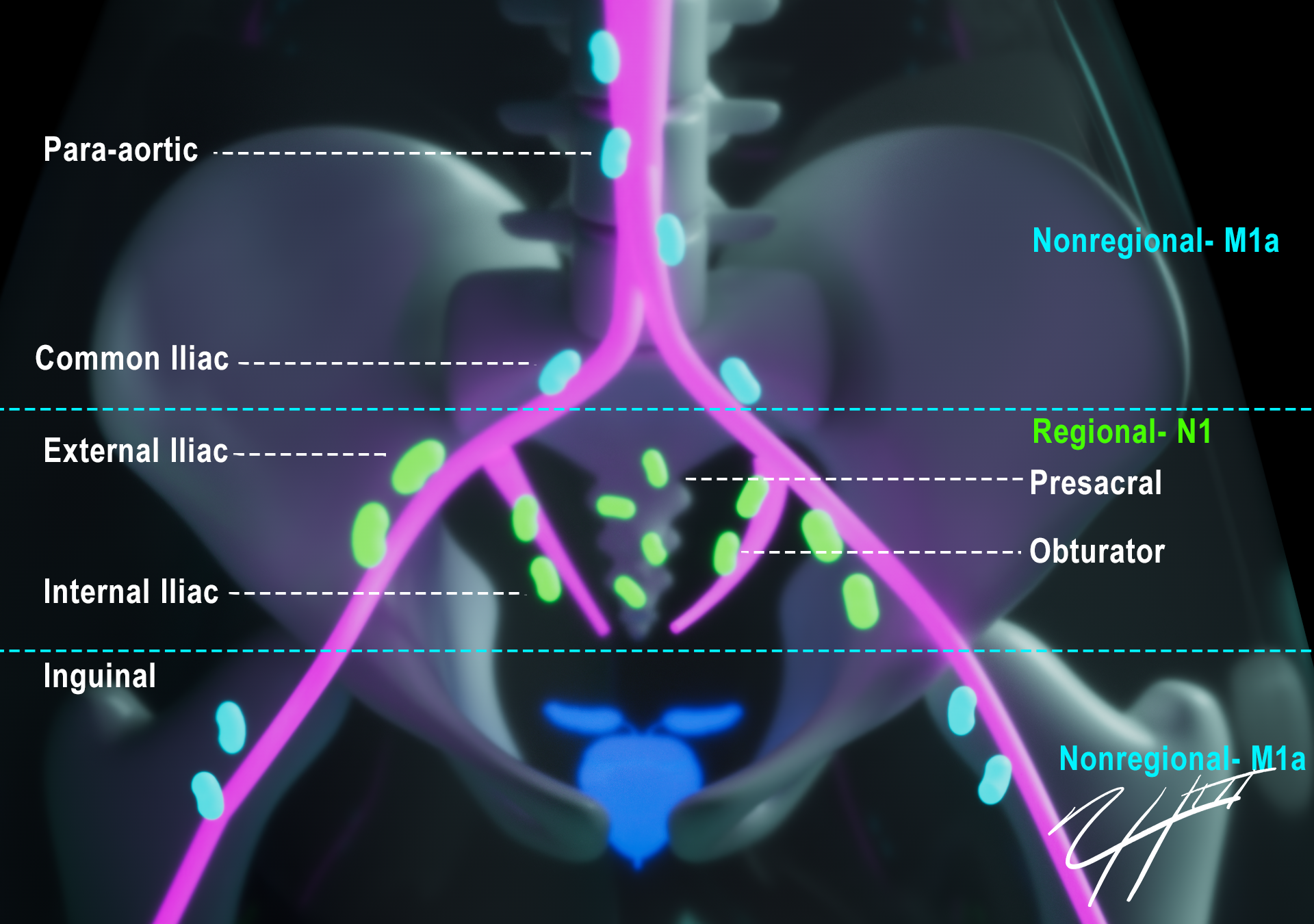
Where N1 Ends and M1a Begins
Explore the major nodal stations relevant to prostate cancer staging.
Hover over a nodal station to explore it.Tap a nodal station to explore it.
No regional nodal metastasis
No suspicious regional nodes identified.
Regional pelvic lymph-node metastasis
One or more regional nodes. Number and laterality do not change the N category.
- External iliac
- Internal iliac
- Obturator
- Presacral / sacral
- Periprostatic
Nonregional lymph-node metastasis
- Common iliac
- Para-aortic / retroperitoneal
- Inguinal
Find Them on MRI
Scrub through the pelvis to connect the nodal map with axial MRI anatomy.
When Is a Node Suspicious?
Nodal size is useful but imperfect. A short-axis diameter greater than approximately 8 mm is suspicious, but metastatic nodes may be normal-sized.
Conventional MRI relies on size and morphology and has limited sensitivity for small-volume nodal metastatic disease. A negative conventional MRI does not exclude microscopic nodal metastases.
Report What Matters
Name the station. Measure the short axis. State the nodal station and staging implication.
Sources
- American College of Radiology, European Society of Urogenital Radiology, AdMeTech Foundation. PI-RADS: Prostate Imaging Reporting and Data System, Version 2.1. 2019.
- Brierley JD, Gospodarowicz MK, Wittekind C, eds. TNM Classification of Malignant Tumours, 8th Edition (UICC). Prostate chapter, AJCC Cancer Staging Manual, 8th Edition.
- European Association of Urology (EAU). Prostate Cancer Guidelines.