MRI or PET?
"If PSMA PET can find prostate cancer throughout the body, why do I still need a prostate MRI?"
The short answer is that MRI and PSMA PET measure different things. Prostate MRI provides high-resolution anatomy. It is designed to localize lesions within the gland, evaluate morphology and diffusion characteristics, and assess local tumor extent. PSMA PET maps PSMA expression. Its major strength is identifying disease beyond the prostate, particularly nodal and distant metastatic disease. They are complementary tests, not interchangeable ones.
Why MRI still matters for local staging
When the question is local anatomy, MRI provides much better soft-tissue contrast than CT or PET-based anatomic images. This is why MRI remains critical for evaluating the prostate capsule, seminal vesicles, adjacent organs, and local tumor extent.
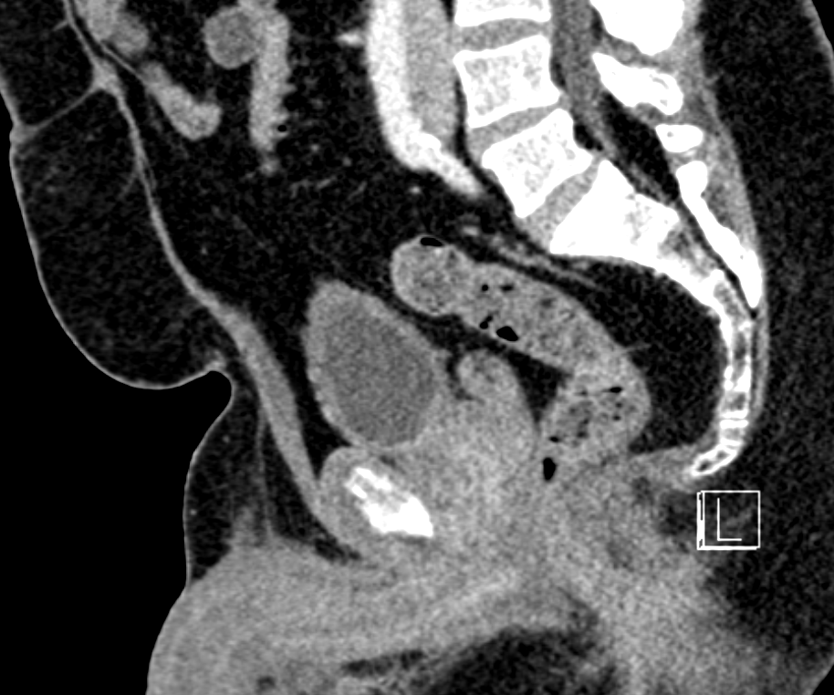
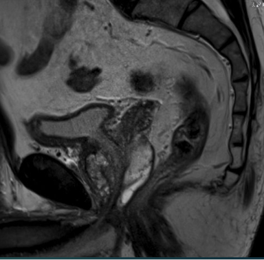
MRI provides substantially better local soft-tissue resolution than CT, making it more useful for intraprostatic localization and assessment of local tumor extent.
Direct comparison
Both tests contribute to prostate cancer diagnosis and staging, but their strengths sit in different places.
| Feature | MRI | PSMA PET |
|---|---|---|
| Detection of clinically significant cancer | Established first-line test. High sensitivity and central to MRI-targeted biopsy pathways. | Promising and complementary. Can detect clinically significant cancer and add information after negative or equivocal MRI, but is not currently a replacement for routine prebiopsy MRI. |
| Localization within the gland | Excellent spatial and anatomic detail. Defines lesion location, margins, and relationship to the capsule, urethra, apex, and adjacent structures. | Strong molecular localization, but lower spatial resolution and dependent on PSMA expression. Small or low-uptake tumors may be occult. |
| Local staging: EPE / SVI | Major strength. Provides detailed morphology of the capsule, neurovascular bundles, seminal vesicles, and adjacent organs. | Can contribute to local staging, but PET uptake does not provide the same detailed anatomic information. Hybrid PET/MRI may help combine these strengths. |
| Nodal and distant staging | Limited for small nodal metastases. Morphologically normal nodes may contain tumor. | Major strength. More sensitive than conventional MRI or CT for nodal staging and provides whole-body metastatic assessment. |
| Important blind spot | Some clinically significant tumors are MRI occult. | Some prostate cancers show low or heterogeneous PSMA expression. PSMA uptake is not equivalent to PI-RADS or tumor grade. |
PSMA uptake is not PI-RADS
A paired case makes this concept concrete. The same gland contained two separate biopsy-proven lesions in the midgland with opposite imaging behavior.
Left midgland: PSMA-avid lesion. Biopsy: Gleason 3+3.
Right midgland: MRI-visible lesion. Biopsy: Gleason 4+3. Less conspicuous PSMA uptake.
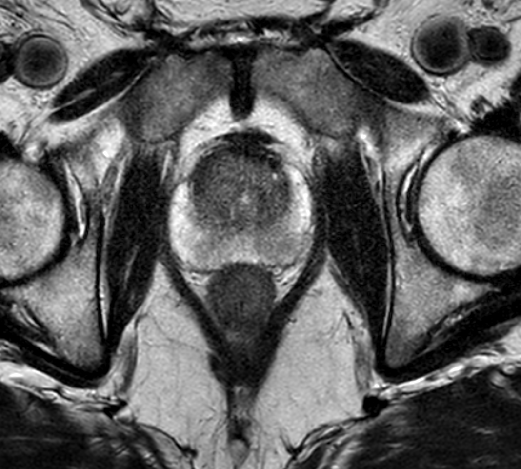
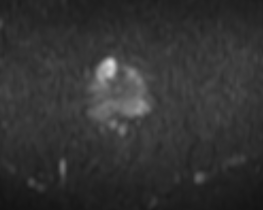
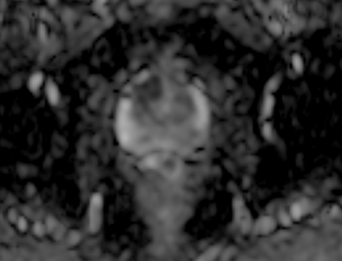
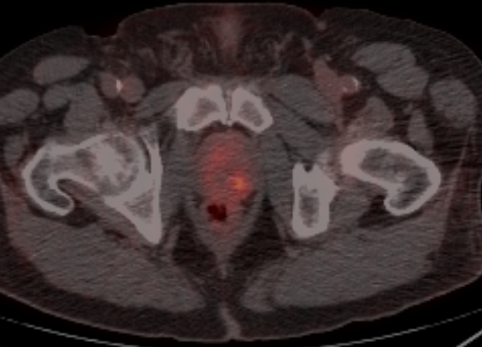
Higher PSMA uptake is associated with more aggressive prostate cancer at a population level, but uptake in an individual lesion does not reliably identify which tumor focus contains the highest-grade disease.
PSMA expression and MRI suspicion reflect different aspects of tumor biology.
Why might a prostate cancer not be PSMA-avid?
PET partial-volume effects can reduce conspicuity.
Different portions of the same tumor may express different amounts of PSMA.
Tumor intermixed with benign tissue may produce less concentrated tracer uptake.
PSMA expression varies among tumors and among different foci within the same prostate.
Where PSMA PET changes the game
Conventional MRI and CT rely heavily on lymph-node morphology and size. Microscopic metastatic disease may therefore exist in a normal-sized node. PSMA PET assesses molecular expression rather than simply nodal enlargement.
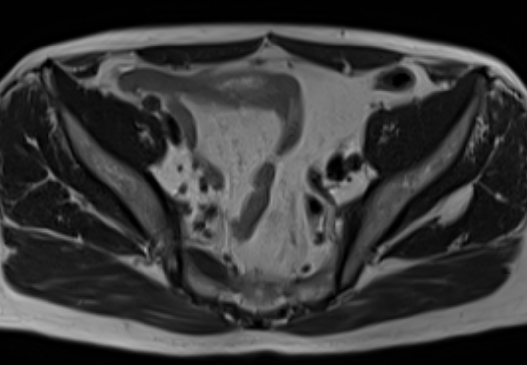
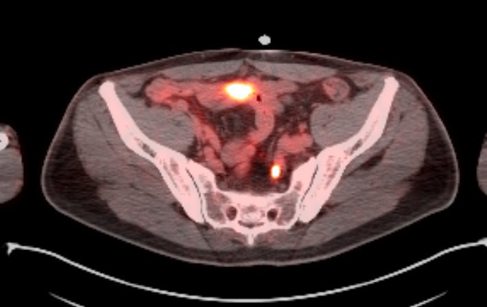
The left internal iliac lymph node is morphologically normal on MRI but demonstrates focal uptake on PSMA PET.
What about PSMA PET/MRI?
Potentially the best of both worlds?
Hybrid PSMA PET/MRI combines MRI's high-resolution anatomy and diffusion imaging with PSMA PET's molecular information. Early studies suggest that combining PET and MRI may improve intraprostatic tumor detection and provide complementary staging information.
However, PET/MRI remains less widely available, more expensive, and operationally more complex than prostate MRI or PSMA PET/CT. For most patients today, separately acquired prostate MRI and PSMA PET/CT provide complementary information.
The emerging question may not be MRI OR PET, but when PET adds useful information to MRI.
Quick answer
| Clinical question | Best imaging strength |
|---|---|
| Is there a suspicious intraprostatic lesion? | MRI |
| Where should I target a biopsy? | MRI, with PSMA PET potentially complementary |
| Is there extraprostatic extension? | MRI for detailed local anatomy |
| Is there seminal vesicle invasion? | MRI, with complementary PET information possible |
| Are pelvic lymph nodes involved? | PSMA PET |
| Is there distant metastatic disease? | PSMA PET |
| MRI is negative or equivocal but clinical concern remains? | PSMA PET may add value in selected patients |
| Which lesion is highest grade? | Do not assume the most PSMA-avid lesion is the highest-grade lesion. |
Prostate MRI remains critical for intraprostatic localization and local staging because of its spatial resolution and soft-tissue characterization.
PSMA PET excels at nodal and distant staging but depends on tumor PSMA expression and should not be interpreted as a molecular PI-RADS score.
The modalities are complementary, not competitors.
References and further reading
- Lemos A, et al. Not always a perfect match: discrepancies between PSMA PET/CT and MRI in prostate cancer diagnosis and staging. Abdominal Radiology. 2026.
- Hofman MS, et al. proPSMA trial. Lancet. 2020.
- Buteau JP, et al. PRIMARY2. Lancet Oncology. 2026.
- PRIMARY study.
- Histopathologic correlation of PSMA-negative/heterogeneous primary prostate tumors.
- PSMA PET/MRI systematic review and meta-analysis.
- EAU Prostate Cancer Guidelines. Diagnostic Evaluation.
Related tools and articles
Written by Nicholas H. Shaheen, MD • Last reviewed August 2026